Document Actions
Retina CAD
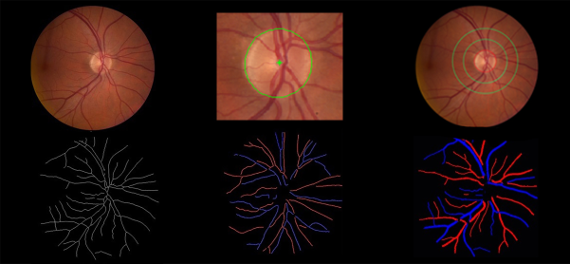
Design of advanced methodologies for early detection of retinal pathologies, based on the automatic calculation of specific markers. These markers are obtained in different eye image modalities (retinography, angiography, Optical Coherence Tomography – OCT).
The development of image processing and analysis methods to extract quantitative information from retinal images has been an active area of research for several decades. Automated detection of lesions or vessel changes from fundus photographs can provide useful information for detecting eye diseases in early stages. Although the greatest emphasis in automated diagnosis has been given to the detection of diabetic retinopathy, image analysis methods are used for a reliable assessment of vascular changes in retinal images as an aid for the diagnosis of the ocular manifestations of a variety of worldwide major diseases such as diabetes, hypertension and the risk of cardiovascular disorders.
Nevertheless, a major limitation of fundus photography is the fact that it is a 2D projection of the 3D retinal surface and underlying tissues. The application of Optical Coherence Tomography (OCT) to retinal imaging has allowed a real 3D sectioning of the retina, making it possible to have high-quality, cross-sectional images of the macula or optic nerve head (ONH). As OCT is recent when compared to fundus photography, the use of image analysis in OCT images is still a new and promising area of research.
At the Biomedical Imaging Lab we want to develop image analysis methodologies for extracting clinically relevant information both from fundus photographs and OCT images of the retina aiming at implementation of CAD systems devoted to the early diagnosis and follow up of eye diseases and systemic diseases with ocular manifestations.
People/Institutions
Behdad Dashtbozorg (INEB), Raul Pinheiro (INEB), Ana Maria Mendonça (INEB), Susana Penas (CHSJ), and Aurélio Campilho (INESC TEC).
Funding
Fundação para a Ciência e a Tecnologia (FCT) and Instituto de Engenharia Biomédica (INEB).
Main publications
- Behdad Dashtbozorg, Ana Maria Mendonça, Aurélio J. C. Campilho:
An Automatic Graph-Based Approach for Artery/Vein Classification in
Retinal Images. IEEE Transactions on Image Processing 23(3): 1073-1083
(2014). Link
- Ana
Maria Mendonça, António V. Sousa, Luís Mendonça, Aurélio J. C.
Campilho: Automatic localization of the optic disc by combining vascular
and intensity information. Comp. Med. Imag. and Graph. 37(5-6): 409-417
(2013). Link
- Ana
Maria Mendonça, Aurélio C. Campilho: Segmentation of retinal blood
vessels by combining the detection of centerlines and morphological
reconstruction. IEEE Trans. Med. Imaging 25(9): 1200-1213 (2006). Link
PhD. Theses
- Behdad Dashtbozorg, Advanced Image Analysis for the Assessment of Retinal Vascular Changes. Doctoral Degree on Electrical and Computer Engineering, FEUP, (Supervisor: Ana Maria Mendonça, co-supervisor: Aurélio Campilho). (ongoing).
- Raul de Medina Prata Pinheiro,
Advanced methods for optical coherence tomography choroidal image
analysis. Doctoral Degree on Electrical and Computer Engineering, FEUP,
(Supervisor: Ana Maria Mendonça). (Ongoing).
- Ana Maria
Rodrigues Sousa Faria de Mendonça, Métodos de Tratamento Digital de
Imagem. Aplicação no Arquivo, Processamento e Análise de Imagens de
Oftalmologia. Doctoral Degree on Electrical and Computer Engineering,
FEUP, (Supervisor: Aurélio Campilho), 1994.
Master Theses
- Filipe José Pinto de Lima Cardoso, Detecção automática de estruturas anatómicas em imagens digitais da retina. Master degree on Electrical and Computer Engineering, FEUP. (Supervisor: Ana Maria Mendonça), 2011.
- Carmen González Pijuan. Alinhamento de Imagens da Retina Usando a Estrutura Vascular e Transformada de Distância. Master in Biomedical Engineering, FEUP. (Supervisor: Ana Maria Mendonça), 2009.
- Raquel Alexandra Afonso Guerra, Identificação automática do disco óptico em imagens coloridas de retina. Master in Biomedical Engineering, FEUP. (Supervisor: Ana Maria Mendonça), 2008.
