Document Actions
Carotid CAD
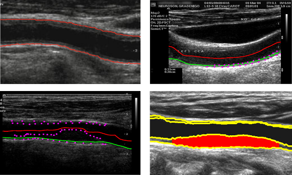
The Carotid CAD project aims at measuring macro vascular markers of atherosclerosis, such as the intima-media thickness (IMT) and plaque burden from carotid US images. The relation of these markers to the presence of cerebral vessel diseases will be studied.
Ultrasonography of the common carotid artery (CCA) has become widely used in clinical practice for the diagnosis of atherosclerosis and associated diseases, as it is a high resolution, noninvasive, low cost and readily available medical imaging technology. From a longitudinal B-mode ultrasound image of the CCA, it is possible to measure the intima-media thickness (IMT), the lumen diameter, or to identify atherosclerotic plaques.
The measurement of macro vascular markers related to the intima-media thickness, the degree of stenosis, the plaque burden or plaque morphology from these ultrasound images of the carotid can be used for early diagnosis of atherosclerosis and stroke risk stratification.

The main objective of the Biomedical Imaging Lab consists of designing and implementing methodologies to automatically obtain these vascular markers and develop CAD tools that help the expert clinicians to early diagnose the patients’ condition.
People/Institutions
José Rouco (INESC TEC), Miguel Duarte (INESC TEC), Rui Rocha (INESC TEC), Jorge Silva (INESC TEC), Elsa Azevedo (CHSJ), Carmen Ferreira (CHSJ) and Aurélio Campilho (INESC TEC).
CHSJ – Centro Hospitalar São João.
Funding
Fundação para a Ciência e a Tecnologia (FCT) and Instituto de Engenharia Biomédica (INEB).
Main publications
- Rui Rocha, Jorge Silva and Aurélio Campilho, Automatic detection of the carotid lumen axis in B-mode ultrasound images, Computer Methods and Programs in Biomedicine, 115 (2014), 110-118. Link
- José Rouco, Aurélio J. C. Campilho: Robust common carotid artery lumen detection in B-mode ultrasound images using local phase symmetry. ICASSP 2013: 929-933. Link
- Rui Rocha, Jorge Silva, Aurélio Campilho, “Automatic segmentation of carotid B-mode images using fuzzy classification”, Medical & Biological Engineering & Computing, Vol. 50, Issue 5 (2012), 533-545. Link
- Rui Rocha, Aurélio Campilho, Jorge Silva, Elsa Azevedo and Rosa Santos, “Segmentation of Ultrasound Images of the Carotid using RANSAC and Cubic Splines”, Computer Methods and Programs in Biomedicine, 101 (2011), 94-106. Link
- Rui Rocha, Aurélio Campilho, Jorge Silva, Elsa Azevedo and Rosa Santos, “Segmentation of the Carotid Intima Media Region in B-mode Images”, Image and Vision Computing 28 (2010), 614-625. Link
PhD. Thesis
- Rui António Henrique Fernandes da Rocha, Image Segmentation and Reconstruction of 3D Surfaces from Carotid Ultrasound Images, Doctoral Degree on Electrical and Computer Engineering, FEUP, (Supervisor: Aurélio Campilho; Co-supervisor: Jorge Alves Silva), 2008.
Master Theses
- Miguel Matos Fernandes de Oliveira Duarte. Automatic measurement of atherosclerotic plaque burden in ultrasound images of the carotid artery. Master in bioengineering, FEUP. (Supervisors: José Rouco and Aurélio Campilho). 2014.
- Classification Approach for Diagnosis of Arteriosclerosis Using B-mode Ultrasound Carotid Images. Catarina de Brito Carvalho. Master in Biomedical Engineering. (Supervisor: Aurélio Campilho). 2012.
